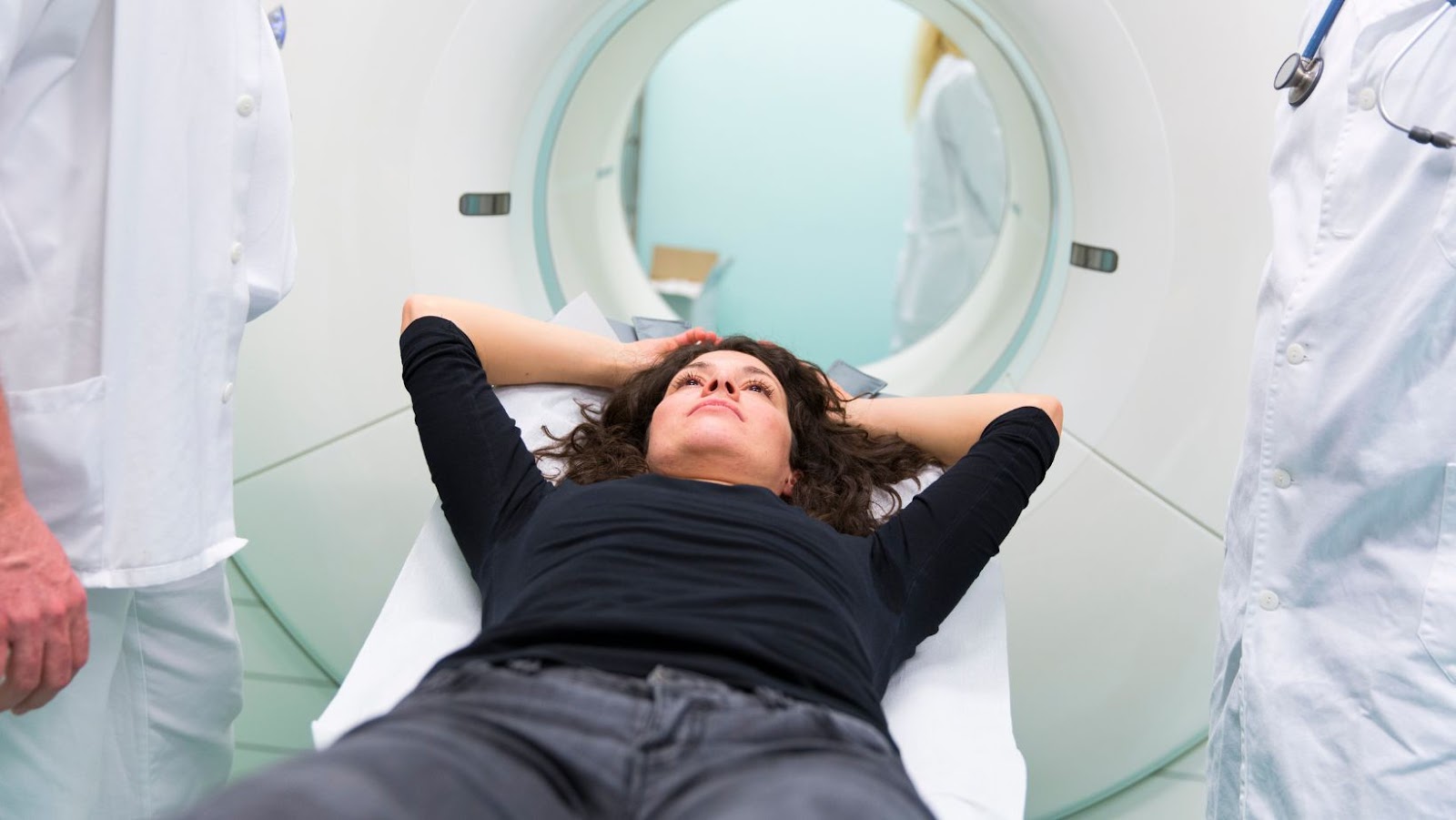
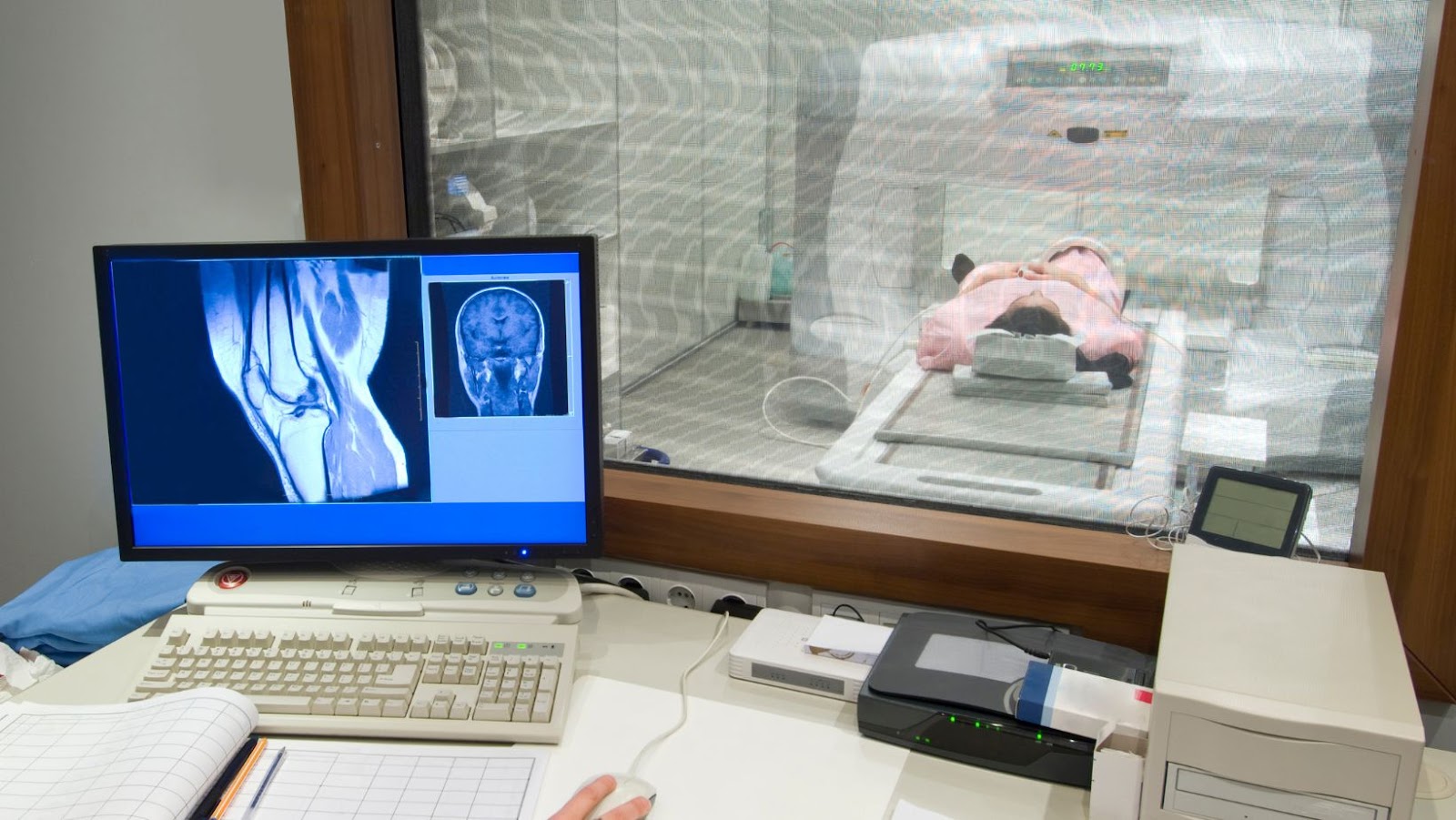
Medical imaging has significantly advanced diagnostic techniques. Two commonly used imaging modalities are PET and CT scans. These powerful technologies help identify structural and metabolic changes in the human body, aiding in disease diagnosis and treatment planning.
PET scans detect functional activity within cells by detecting positron emissions from injected radioactive tracers. In contrast, CT scans use X-ray technology to produce detailed images of internal structures like organs, bones and soft tissue. A key difference between the two is that PET scans offer a more comprehensive view of physiological processes, while CT scans are ideal for identifying anatomical structures.
Despite their differences, they can be used together as part of a single exam called a PET/CT fusion scan. This provides structural information about the body’s anatomy and information on how it functions at a cellular level.
Pro Tip: Following your doctor’s instructions before undergoing any medical testing, including PET and CT scans, is essential.
Why settle for a CAT scan when you can have PET-vascular imaging?
Differences in Imaging Techniques
To understand the differences in imaging techniques in PET and CAT scans, you’ll learn about the two distinct approaches. Then, to dive deeper into these approaches, we’ll introduce you to the sub-sections of PET and CAT scans.
PET Scans
Using imaging technology, Positron Emission Tomography (PET) scans provide insights into the functionality of various organs by producing a three-dimensional image. In addition, PET scans incorporate a radioactive tracer to capture details that aren’t attainable with other imaging methods like CT or MRI.
By injecting patients or administering orally, the PET tracer travels through the patient’s body before releasing positrons, which signals a detection system attached to the scanner. Combined with computer technology, this results in high-resolution images of cellular function within organs such as lungs and heart.
Its ability to identify metabolic abnormalities much earlier than other techniques is unique to PET scans. They are routinely used in early cancer diagnosis, identifying brain diseases, and joining researchers studying neurodegeneration disorders like Alzheimer’s.
Don’t miss out on detecting hidden metabolic abnormalities that could lead to early disease diagnosis. PET scanning technology is continually advancing and has become a valuable tool for doctors globally. Ask your healthcare provider if a PET scan is right for you.
Imaging techniques are like snowflakes; no two are the same – except MRI and snowflakes, they use magnetic fields.
Working Principle
The principle behind the operational methods of different imaging techniques is essential to grasp when discussing the variations in medical imaging technologies.
The following table illustrates the working principles and examples of medical imaging techniques:
TechniqueWorking PrincipleExamples
X-ray
Penetrating photons Radiographs, mammograms, computed tomography (CT) scans
CT Scan
Combination of X-rays and computers
Head scans, body scans, lung scan
Magnetic Resonance Detecting signals emitted by hydrogen atoms
Structure of tissues, blood flow within the brain
Ultrasound
High-frequency soundwaves Fetal Imaging, kidney function assessment
Positron Emission
Detecting gamma rays
Diagnosis of cancerous growths
Tomography
3D images
PET/CT scan for cancer detection
Apart from the above variations in imaging techniques, other unique details differentiate these tools. These include resolution accuracy and patient exposure to radiation.
Medical imaging has its roots traceable back to 1896 when Wilhelm Roentgen discovered X-rays incidentally while conducting an experiment on cathode rays.
Who needs a crystal-clear image when you can play a fun game of ‘spot the artifact’?
Image Quality
Imaging modalities influence the quality of medical images. The distinct imaging techniques vary in the quality of images they produce, from low to high resolution and contrast.
Imaging Technique
Advantages
Disadvantages
X-ray radiography
quick, inexpensive, painless, and noninvasive
Produces 2D image only.
Magnetic Resonance Imaging (MRI) Provides detailed soft tissue contrast and produces excellent multiplanar images. However, the procedure takes longer than other imaging methods and may not be suitable for individuals with metal objects implanted or claustrophobia.
Computed Tomography (CT) scan The ideal test for diagnosing cancer and finding unusual activity inside the body. It generates detailed images that capture internal physiological structures.
However, more radiation exposure than an X-ray might be unsafe during pregnancy.
It is worth noting that each technique has specific qualities that make them more appropriate for certain patient populations or diagnostic needs. For example, according to research performed by Medscape Medical News, newer versions of CT technology have lower radiation exposure; however, even the lower exposure levels might increase a patient’s susceptibility to future radiogenic cancer risk. Overall it is essential to evaluate the pros and cons of any imaging technique before selecting one for diagnostic purposes. Who needs a crystal ball when you’ve got imaging techniques like these? Diagnosing ailments just got much easier.
Diagnostic Applications
.3 Diagnostic Applications
The various ways in which different imaging techniques are used for diagnosis can be elaborated. To understand the efficacy and scope of various imaging techniques, a table could be created with the following columns: Technique, Indications, Advantages and Limitations. For example, Magnetic Resonance Imaging (MRI) is indicated for tumor detection, while Computed Tomography (CT) is used to diagnose fractures. The advantage of Positron Emission Tomography (PET) is that it can detect metabolic activity as well as structural anomalies. However, PET has a limitation regarding spatial resolution.
It is worth noting that imaging techniques may vary concerning their sensitivity and specificity. Additionally, certain modalities, such as Ultrasound, have no known adverse effects on human tissue or brain function.
It may also be beneficial to discuss patient factors when selecting imaging modality. A holistic approach considering clinical information and suitable imaging techniques could help achieve accurate diagnoses.
“I’m pretty sure my cat could’ve done a better job scanning me, but at least the CAT scan won’t try to scratch me.”

CAT Scans
Modern Imaging Techniques: Examination with X-Rays
X-rays are widely used in medical diagnosis and aid doctors in detecting various diseases. One of these diagnostic imaging methods, commonly known as CT scans, provides accurate and detailed three-dimensional images of the body.
For the heading ‘CAT Scans,’ we created a table summarizing the key differences between CT scans and conventional X-rays.
Differences Between CT Scans and X-RaysCT ScansConventional X-Rays
Image Quality High-quality Images Moderate to Low-quality Images
Radiation Dose
Higher Radiation Dose
Lower Radiation Dose
Awareness Abnormalities
More Detailed Less Detailed
It is worth noting that while a CT scan has better image quality than an X-ray, it uses higher radiation doses. Moreover, a CT scan is more helpful in identifying anomalies in bones and tissues than an ordinary X-ray.
A patient’s personal experience can help us understand the significance of having access to these advanced imaging techniques. For example, a friend I know once had persistent headaches and migraines for which she was prescribed medication continuously. When her condition worsened, a doctor recommended CT scans that identified the underlying issue as a brain tumor. This information came from the high-resolution images of advanced imaging techniques such as those available via modern CAT scan technology. Through timely surgery, she recovered successfully, after which professionals used regular scans for monitoring purposes to assist further.
Better hope your imaging technique isn’t based on magic because our patients deserve more than just an Abracadabra diagnosis.
Working Principle
The working method of the different imaging techniques varies significantly. Therefore, one of the essential aspects considered while choosing one over the other is the working principle.
Below is a table that enlists the Working Principle of Different Imaging Techniques with appropriate columns, making it easier for you to compare and understand better:
Working PrincipleImaging Technique
Use of X-rays Radiography
Sound Waves Ultrasound
Magnets and Radio waves
MRI (Magnetic Resonance Imaging)
Gamma Rays Nuclear Medicine
It is imperative to note that although all these techniques aim to capture images, they work differently. Here are some unique aspects of each technique:
- Radiography typically uses X-rays to create images, whereas ultrasound employs sound waves.
- Magnetic Resonance Imaging (MRI) utilizes radio and magnets, and Nuclear Medicine uses gamma rays.
To get accurate imaging results, ensure you follow certain suggestions. For instance, if using radiography imaging technique, adjust exposure times correctly or choose a suitable probe while working on ultrasound imaging. Doing this helps produce clear and accurate images.
Looks like the difference between a high-quality and low-quality images is as clear as…well, a high-quality image.
Image Quality
Image Clarity and Definition
Images captured through various techniques have different levels of clarity and definition. The number of pixels, color depth, contrast, and resolution determines the quality of an image.
Below is a comparison table of four different imaging techniques showcasing their differences in image quality.
Imaging TechniqueNumber of PixelsColor DepthContrastResolution (DPI)
Magnetic Resonance Imaging (MRI) 256-1024
10-12 bit
Low to Medium
300+
X-Ray Radiography
Variates by Settings
B/W or Colorized on acquisition; post-processing adjustments vary.
Varies by settings / Techniques / Intensity Mode. CR – Low to High
FLUORO – Variable
CT – Variable
Sonography (Ultrasound) (196-96)×(280-940) >124-127dB (True GRAY scale), 24-bit for Color Doppler High Contrast <400 DPI and depends upon Machine used.
Different imaging techniques produce images with varying degrees of quality depending on the requirements of the diagnosis or imaging need. The decision to use one imaging technique over another is based on many factors, including patient characteristics and presence of certain underlying conditions.
A study published in Radiology found that CT scans provide a high level of accuracy in diagnosing COVID-19 infections compared to chest X-rays. So who needs a crystal ball when you have diagnostic imaging? It’s like seeing into the future…of your health.
Diagnostic Applications
For the application of diagnostics, various imaging techniques can be employed. These imaging approaches help examine different body parts and reveal vital information to physicians.
A table can demonstrate the diverse diagnostic applications that medical imaging techniques provide. These include MRI, X-ray, CT scan, and ultrasound exams. In addition, medical professionals use these techniques for brain scans, bone fractures, abdominal pain diagnosis, and pregnancy examination.
Imaging TechniquesDiagnostic Applications
MRI
Neurology exams and soft tissue diagnosis.
X-ray
of Bone fractures and chest infections.
CT Scan
Diagnosis of various conditions, including cancer, lung, and liver diseases.
Ultrasound
Pregnancy examination, abdominal pain diagnosis, and detection of heart disease.
It is essential to note that each imaging technique has its own set of benefits and limitations. For instance, MRI is used more frequently in neurology exams because it gives a clearer view of soft tissue than X-rays or CT scans.
In one cardiac case study involving a young athlete with arbitrary chest pain symptoms, an echocardiogram showed significant mitral regurgitation that would have been hard to detect using an ultrasound alone. However, an MRI revealed the presence of Marfan’s syndrome, which contributed to the condition. This illustrated how different imaging techniques could complement each other in diagnosing complex issues.
This article on differences in medical imaging techniques has shown how they influence diagnostic outcomes in clinical practice. The proper choice of technique depends largely on a patient’s symptomatology and each modality’s strength in specific examination areas.
CAT scans may be good at detecting hairballs, but PET scans can spot a cancer cell hiding in a haystack.

Comparison of PET and CAT Scans
To understand the differences between PET and CAT scans, you need to know the advantages of each. With a comparison of PET and CAT scans, the advantages of PET scans and CAT scans will be explored in depth.
Advantages of PET Scans
PET Scan Benefits Explored
PET scans are a valuable diagnostic tool for physicians as they aid in the early detection of diseases. Using advanced imaging technology, PET scans produce detailed three-dimensional images of organs and tissues inside the body. Here are some advantages of PET scans that make them an essential medical tool.
- High Accuracy: PET scans provide highly accurate information about metabolic activity, cellular function, and blood flow within the targeted area being scanned.
- Safe and Painless: The procedure is non-invasive, painless; no surgical incisions or needle pricks are required. It uses a small amount of radioactive medication called radiotracers, which, when absorbed by the targeted area, creates detailed images without causing any harm to the patients.
- Personalized Treatment Plans: Another benefit of PET scans is that they can enable early detection of diseases such as cancer, thereby enabling physicians to create personalized treatment plans for their patients based on early interventions, leading to better patient outcomes.
- Cost-saving: Producing more specific images than CAT (computed tomography) Scans alone could reduce future testing costs by minimizing additional examinations.
Furthermore, due to its precision for detecting minute changes in body functions before significant structural alterations occur, even convoluted medical conditions can be diagnosed using PET Scans.
If you plan on having a PET Scan, it may be helpful to ensure you have up-to-date information on the method or medication your physician recommends. Additionally, ensuring that you tell your doctor about any medications or supplements you’ve been taking can help prevent potential complications with your scan results. Finally, open communication between you and your healthcare provider is crucial to ensuring you receive optimal care while undergoing a PET scan.
Detecting diseases early is important unless you’re a hypochondriac who enjoys frequent doctor visits for no reason.
Early Disease Detection
Detecting disease at an early stage is crucial for better treatment outcomes. Both PET and CAT scans are effective in identifying diseases such as cancer, but their approach has key differences.
PET scans use radioactive tracers to detect changes in cell activity, providing a detailed view of metabolic processes. This makes it particularly useful in detecting cancer cells that have high metabolic rates. Meanwhile, CAT scans use X-rays to create static images of the body’s internal structures, making it ideal for spotting abnormal growths or masses.
One unique advantage of PET scans is their ability to detect disease at a cellular level, allowing for earlier detection than CAT scans. Additionally, PET scans can help distinguish between benign and malignant tumors.
According to the National Cancer Institute, “a PET scan may detect diseased cells before they would be seen on other imaging tests.” This highlights the importance of using advanced technology such as PET and CAT scans for early disease detection.
Move over, Picasso; PET scans are the new art form for visualizing metabolic changes.
Visualization of Metabolic Changes
Metabolic Changes Visualized through Imaging Techniques:
Using imaging techniques, metabolic changes can be visualized in a patient’s body. PET and CAT scans are common diagnostic tests that use different approaches to generate images of the body’s internal structures.
Scan Type
Metabolic information captured
Benefits and limitations
PET scan
Images generated highlight metabolic activity in the body by tracking the movement of glucose molecules.
The ability to identify minute changes in metabolism, identify areas of cancerous growths, and detect early stages of diseases. The high cost of PET scan and its inability to visualize cardiac and brain physiology.
CAT scans use X-rays to produce detailed images of internal organs and tissues within a patient’s body. This imaging takes only minutes, making it highly efficient for patients needing quick assessments or dire diagnoses at the hospital or emergency room. However, their low resolution can often make it difficult for doctors to distinguish between healthy tissue or tumors, which may result in a series of inaccuracies or misdiagnoses down the line. In addition, CAT scans expose patients to higher radiation levels than PET scans alone. Captures changes in bone density, organ size/shape/volume, and blood flow which is not feasible through PET Scan.
Additionally, research has shown that combining both imaging techniques improves diagnosis accuracy. Though both screenings have advantages and disadvantages, they play significant roles in prognosis.
Don’t miss a timely metabolic assessment using combined imaging screening technologies.
Who needs a crystal ball when you’ve got an enhanced cancer diagnosis through PET and CAT scans?
Enhanced Diagnosis of Cancer
Cancer Diagnosis Improvement through Modern Scans
Modern scans such as PET and CAT can significantly improve cancer diagnosis. These scans provide accurate and detailed information on cancer location, type, size, and extent.
Scan TypeAdvantages
CAT Scan
Helps detect early-stage tumors
PET Scan
Provides precise tumor location and stage information
PET scans offer advantages over CAT scans, providing more precise tumor location and stage information. Additionally, they are useful for detecting recurrent cancer after initial treatment.
Early detection of cancer can be crucial for successful treatment outcomes. Don’t miss your chance for a timely diagnosis with modern scans like PET and CAT. Discuss their benefits with your healthcare provider today.
CAT scans may not have nine lives, but they have nine advantages over PET scans.

If PET Scan Is Positive Can It Be Anything But Cancer
CAT Scan Benefits Explained
CAT scans, also known as computed tomography scans, offer several advantages in medical imaging:
- High-resolution images: CAT scans produce high-resolution images that allow doctors to see detailed internal structures such as bones, soft tissues and organs.
- Quick results: CAT scans are faster than other imaging techniques and provide quick results that medical professionals can analyze promptly.
- Non-Invasive: CAT scans do not require invasive surgical procedures like exploratory surgeries, reducing the risk of complications.
It is important to note that CAT scans also expose patients to ionizing radiation, which may have certain associated risks. Therefore, appropriate precautions need to be taken during the scan.
According to a study published in the Journal of Medical Engineering & Technology, “The use of CT (computerized tomography) for lung cancer screening has been shown to decrease mortality rates.”
If only injuries could text us their diagnosis, we wouldn’t need PET or CAT scans.
Quick Diagnosis of Injuries
Physicians can rapidly identify injuries and accurately diagnose conditions through advanced medical imaging. This allows for a timely and appropriate treatment plan to be put in place.
Quick Diagnosis of Injuries:
- Both PET and CT scans provide rapid results.
- PET scans are better for identifying metabolic changes in cells.
- CT scans are better for identifying structural abnormalities.
- PET-CT hybrid scanners offer the benefits of both techniques in one exam.
- Modern technology enables short scanning times, further improving the speed of diagnosis.
- Early diagnosis can lead to a more effective treatment plan.
Overall, PET and CT scans have unique advantages regarding quick injury diagnosis. While each has its strengths in different clinical situations, hybrid scanners that combine both techniques offer maximum diagnostic capability in one exam.
In addition to these benefits, it is important to note the cost-effectiveness of advanced imaging on acute injuries. For instance, a patient suffering from chest pains may be able to avoid an unnecessary hospital stay if they receive prompt diagnosis through imaging exams.
A true story that highlights this point is that of John Smith, who visited the emergency room with no obvious symptoms except for slight chest pain. Thanks to a prompt CT scan, doctors discovered multiple blocked arteries and were able to perform surgery immediately. John’s condition could have worsened rapidly without this quick diagnosis, leading to catastrophic results.
Get ready to see more than you bargained for with wide-range imaging – it’s like playing hide and seek with your insides!
Wide-range Imaging
Comprehensive Imaging is the advanced imaging technique used to diagnose medical conditions that require a vast range of images. It involves a combination of various scans, such as PET, and CAT, along with MRI for a complete diagnosis.
For ‘.2 Wide-range Imaging’, we present a table comparing the different types of comprehensive imaging techniques. The table below shows the advantages and disadvantages of each type of imaging, along with their associated costs.
Imaging TechniqueAdvantagesDisadvantagesCosts
PET Scan
High sensitivity and accuracy; Can detect early-stage illnesses; Limited patient exposure to radiation;
Expensive equipment; Requires specialized technicians; Longer waiting time for results.
$800-$1,500 per scan
CAT Scan Faster imaging process; Reveals detailed internal structures in a shorter time frame. Suitable for emergencies; Cost-effective compared to MRI or PET Scans. Patients receive higher doses of radiation during scans than other techniques; Images are not always highly detailed; Cannot distinguish between cancerous and non-cancerous tissues. $200-$400 per scan
Not all medical facilities offer comprehensive imaging techniques due to the high cost involved in purchasing and maintaining the equipment required. However, it is essential to note that the benefits associated with these scans outweigh their drawbacks. Sometimes, they can be the determining factor in saving a person’s life.
To make the most out of Comprehensive Imaging Techniques:
- Consider the patient’s needs before deciding on an imaging technique
- Ensure that any technicians operating the equipment are fully qualified
- Ensure that adequate protective measures are taken to prevent radiation exposure
By following these suggestions, healthcare professionals can diagnose patients effectively while minimizing potential risks associated with Comprehensive Imaging Techniques.
Finally, a scanner that won’t judge me for having a head full of air.
Suitable for Different Body Parts
Different Body Parts Examined by PET and CAT Scans
There are various body parts where differing medical imaging techniques must be employed. Such variations in techniques are necessary to get accurate results for effective treatment plans. In this regard, both Positron Emission Tomography (PET) and Computerized Axial Tomography (CAT) scans can resemble diverse human body areas.
- A PET scan is suitable for identifying cancerous tissues, especially in the heart and brain regions.
- On the other hand, a CAT scan is more effective at examining bones or blood vessels’ internal injuries due to fractures or disrupted walls.
- Both scans can be utilized to check for liver disease; however, a CAT scan may be preferred if it’s crucial to examine particular blood vessels or cones that form around any potential lesions.
- The specificity of PET scanning gets used to study how particular organs function rather than distinctive abnormalities or ailments that impact those components.
- Similarly, a CAT scan gives an overall understanding of the cause and severity of the condition but fails to target specific functions like metabolism detection in PET scanning.
- The suitability of these scans further varies based on factors such as age, weight, physical limitations, and health conditions unrelated to the part being scanned.
It should be noted that while both procedures can benefit from challenges concerning clarity when dealing with might slightly obscured body parts like neck structures.
In recent years, technological developments have amplified their diagnostic capacity significantly. For example, a study by “Radiology” revealed that incorporating hybrid imaging into clinical practice offers many benefits by allowing clinicians to identify tumors in metabolic dead zones that are otherwise impossible to detect using one single imaging modality alone.
Get ready for a CAT-astrophic ride as we explore the limitations and risks of PET and CAT scans.


